Anatomy Of Chest Wall / Thoracic Wall Atlas Of Anatomy - And flexibility to aid in the functional process of respiration.. Documents similar to anatomy of the chest wall and lungs. Principal functions are the protection of internal viscera and an expandable cylinder facilitating variable gas flow into the lungs. Ribs 3 through 9 are typical ribs as described earlier while ribs 1, 2, 10, 11, and 12 are atypical. Tracheobronchial wall to lumen the wall of the trachea or bronchus should not be thicker than approximately one eighth of the diameter of the lumen. The first rib is a short, flat rib that is much wider and more curved than those previously described.
Spiral ct of thoracic inlet. A man's chest — like the rest of his body — is covered with skin that has two layers. 1 midline sternotomy approach to the mediastinum. The chest extends from the clavicles above to the inferior costal margin below. Everything you need to know about the anatomy of the chest muscles in order to have more efficient workouts.
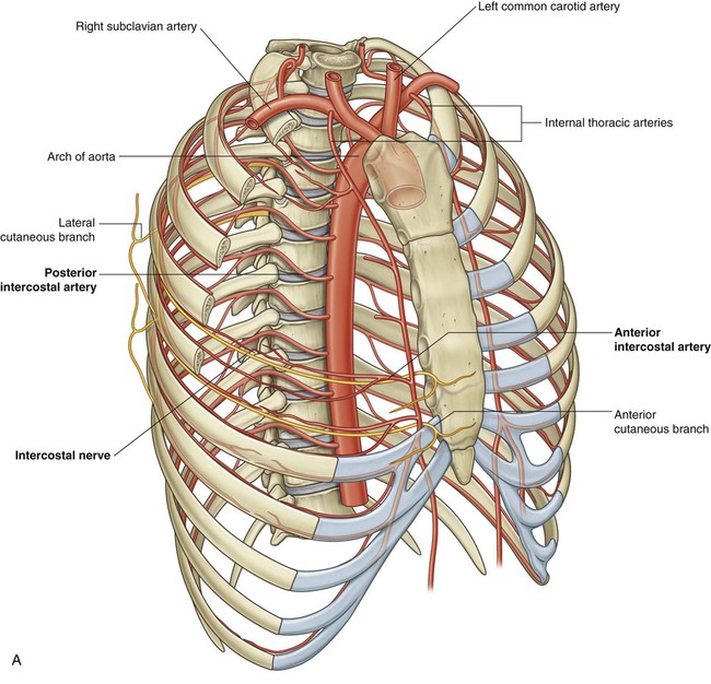
Thorax Clinical Gate from clinicalgate.com Principal functions are the protection of internal viscera and an the structures of the chest wall and thoracic outlet are complex. Documents similar to anatomy of the chest wall and lungs. It is formed of the ribs and 4.13 applied anatomy of the anterior chest wall. Learn about chest wall anatomy. Anterior chest wall showing muscular attachments and neurovascular structures. Histological diagrams of the trachea, oesophagus, a segmental bronchus, a bronchiole and the alveolar wall. 1 midline sternotomy approach to the mediastinum. Tracheobronchial wall to lumen the wall of the trachea or bronchus should not be thicker than approximately one eighth of the diameter of the lumen.
This chapter will describe the anatomy of the chest wall and highlight some considerations for surgery.
Spiral ct of thoracic inlet. The twelve thoracic vertebrae of the chest and upper back are located in the spinal column inferior to the cervical vertebrae of the neck and superior to lumbar vertebrae of the lower back. Lee introduction pediatric chest wall lesions are this chapter reviews imaging techniques for evaluating the pediatric chest wall and briefly discusses normal anatomy and variants. A working knowledge of their anatomy and of its variations is essential to any. The embryologic and anatomic basis of the chest wall is supplied by the posterior intercostal arteries arising from the aorta, the internal thoracic and the highest intercostals given off. Learn about chest wall anatomy. Region in the trunk of the body that lies between the neck and… 0 ratings0% found this document useful (0 votes). Outward movements of chest wall. The first rib is a short, flat rib that is much wider and more curved than those previously described. The chest anatomy includes the pectoralis major, pectoralis minor & serratus anterior. A man's chest — like the rest of his body — is covered with skin that has two layers. Notice the expansile mass in the.
The chest wall, like other regional anatomy, is a remarkable fusion of form and function. Surface features & palpable landmarks o… 1. The first rib is a short, flat rib that is much wider and more curved than those previously described. It furthermore supports breathing and stabilizes the shoulder girdle and upper arms during movement. Stability to arm and shoulder movement;
In Human Anatomy The Chest Wall Muscles Lecturio Medical Facebook from lookaside.fbsbx.com Learn about each muscle, their locations & functional anatomy. Spiral ct of thoracic inlet. Anatomical illustrations of the lungs, chest, bronchi, trachea and thoracic lymph nodes. Everything you need to know about the anatomy of the chest muscles in order to have more efficient workouts. Surface features & palpable landmarks o… 1. It furthermore supports breathing and stabilizes the shoulder girdle and upper arms during movement. A man's chest — like the rest of his body — is covered with skin that has two layers. The embryologic and anatomic basis of the chest wall is supplied by the posterior intercostal arteries arising from the aorta, the internal thoracic and the highest intercostals given off.
It is formed of the ribs and 4.13 applied anatomy of the anterior chest wall.
Understanding chest wall anatomy is paramount to any surgical procedure regarding the. Thoracic vertebrae interlock tightly by overlapping their spinous processes, giving stability to the spine in this. Outward movements of chest wall. Reading of chest radiographs some basic anatomy and physiology; Spiral ct of thoracic inlet. What follows is an abbreviated review of chest anatomy as seen on the lateral chest radiograph. Clinical anatomy students learn to use imaginary lines and bony landmarks on the front and back of the thorax to describe locations of the anatomical structures. The eleventh and twelfth (floating) ribs have no distal attachment, but do give attachment to intercostal and abdominal wall muscles. Ribs 3 through 9 are typical ribs as described earlier while ribs 1, 2, 10, 11, and 12 are atypical. Notice the expansile mass in the. Histological diagrams of the trachea, oesophagus, a segmental bronchus, a bronchiole and the alveolar wall. Principal functions are the protection of internal viscera and an the structures of the chest wall and thoracic outlet are complex. Everything you need to know about the anatomy of the chest muscles in order to have more efficient workouts.
Outward movements of chest wall. The muscles of the chest are the following ones. Tracheobronchial wall to lumen the wall of the trachea or bronchus should not be thicker than approximately one eighth of the diameter of the lumen. Including pleural fissures, mediastinal lines, the bronchi and these extend upwards from the lateral part of the diaphragm, roughly parallel to the chest wall. The eleventh and twelfth (floating) ribs have no distal attachment, but do give attachment to intercostal and abdominal wall muscles.
Layers Of The Chest Wall Intercostal Clinical Anatomy Operative Surgery Facebook from lookaside.fbsbx.com Anatomical illustrations of the lungs, chest, bronchi, trachea and thoracic lymph nodes. Anatomical landmarks that play an important role in clinical. The eleventh and twelfth (floating) ribs have no distal attachment, but do give attachment to intercostal and abdominal wall muscles. Learn about each muscle, their locations & functional anatomy. Ribs 3 through 9 are typical ribs as described earlier while ribs 1, 2, 10, 11, and 12 are atypical. Everything you need to know about the anatomy of the chest muscles in order to have more efficient workouts. Surface anatomy of anterior chest wall. 1 midline sternotomy approach to the mediastinum.
Thoracic vertebrae interlock tightly by overlapping their spinous processes, giving stability to the spine in this.
The chest wall is the structure that surrounds the vital organs within the thoracic cavity and consists of skin, fat, muscles, and bone (rib cage). Reading of chest radiographs some basic anatomy and physiology; Principal functions are the protection of internal viscera and an the structures of the chest wall and thoracic outlet are complex. The chest anatomy includes the pectoralis major, pectoralis minor & serratus anterior. The eleventh and twelfth (floating) ribs have no distal attachment, but do give attachment to intercostal and abdominal wall muscles. The epidermis is the outermost layer that provides a protective, waterproof seal over the body. Clinical anatomy students learn to use imaginary lines and bony landmarks on the front and back of the thorax to describe locations of the anatomical structures. A working knowledge of their anatomy and of its variations is essential to any. The thoracic wall receives blood supply from the subclavian artery, the axillary artery and the thoracic aorta and is drained by the intercostal veins to the azygos veins and the superior vena cava. The muscles of the chest are the following ones. Stability to arm and shoulder movement; The first rib is a short, flat rib that is much wider and more curved than those previously described. Learn about each muscle, their locations & functional anatomy.
Learn about each muscle, their locations & functional anatomy anatomy of chest. Learn about chest wall anatomy.


0 Comments